
What Is a PET CT Scan, and Why Is It Used?
A PET CT scan is a hybrid medical imaging test that combines Positron Emission Tomography (PET) and Computed Tomography (CT) into a single system.
It provides both metabolic (functional) and anatomical (structural) information in one scan session.
In clinical practice, PET CT is used to detect disease at the cellular level before structural changes appear, making it one of the most advanced tools in modern diagnostic imaging.
What Is a PET CT Scan?
A PET CT scanner integrates two imaging technologies:
-
PET (Positron Emission Tomography):
Detects abnormal metabolic activity using radioactive tracers. -
CT (Computed Tomography):
Produces high-resolution cross-sectional images of organs and tissues.
The combined system overlays metabolic data onto precise anatomical images, allowing clinicians to identify, localize, and stage disease accurately.
How Does a PET CT Scan Work?
Step-by-step process
-
Radiotracer injection
Common tracer: 18F-FDG (Fluorodeoxyglucose). -
Uptake period
Patient rests for 45–60 minutes while the tracer distributes in the body. -
PET imaging
Detects gamma photons emitted from positron annihilation events. -
CT imaging
Provides anatomical mapping and attenuation correction. -
Image fusion
PET metabolic signals are fused with CT images for accurate localization.
Key principle:
Cancer cells consume more glucose than normal cells, resulting in higher FDG uptake.
Why Is PET CT Used in Clinical Medicine?
PET CT is primarily used for early detection, staging, therapy assessment, and recurrence monitoring.
Core medical purpose
-
Detect disease before structural damage occurs
-
Differentiate benign vs malignant lesions
-
Measure treatment response accurately
-
Identify residual or recurrent disease
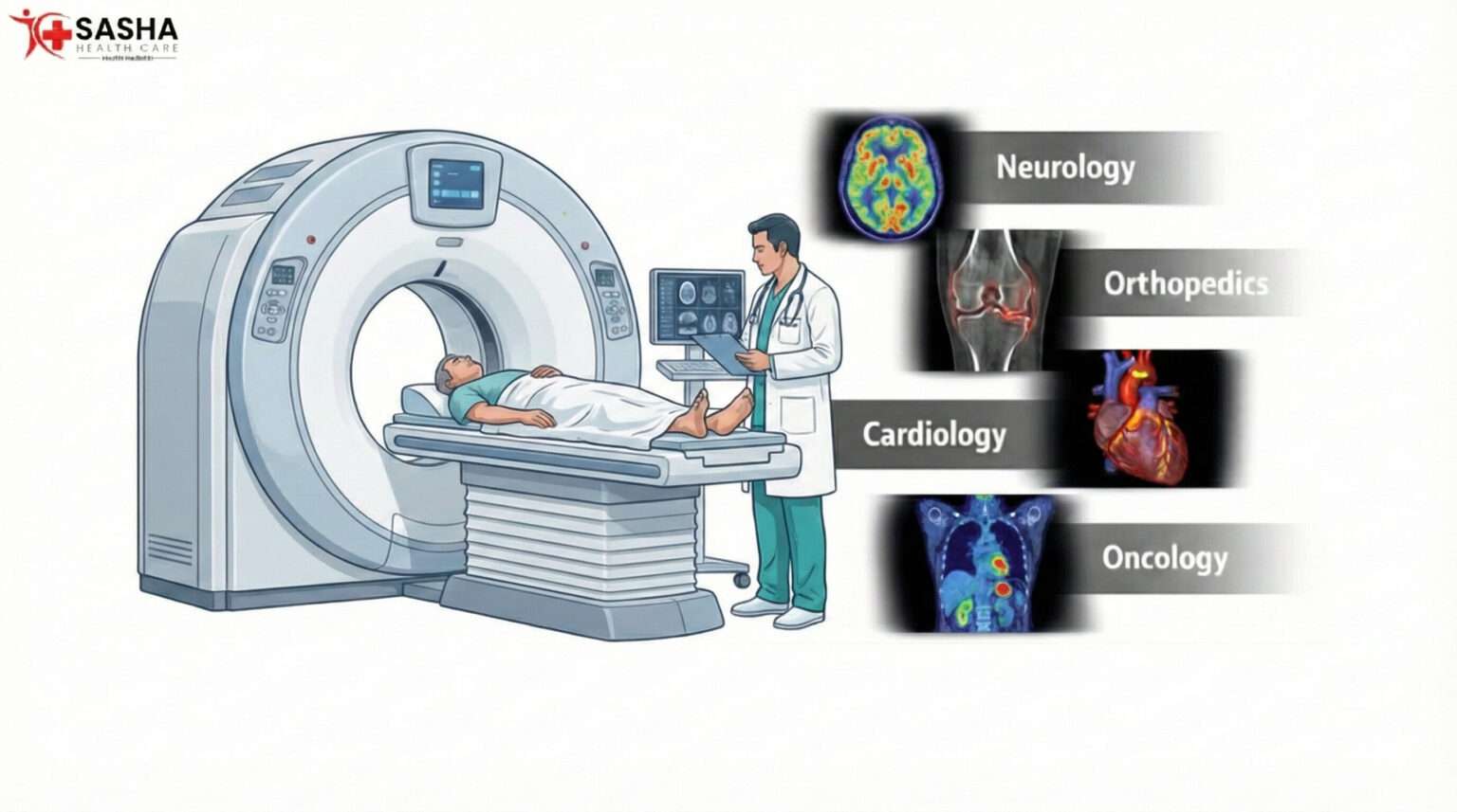
Major Clinical Applications of PET CT
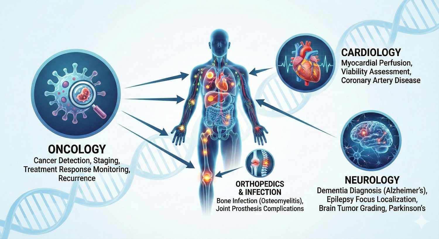
Oncology (Primary Indication)
PET CT is considered the global standard imaging modality in cancer care.
Common cancers evaluated:
-
Lung cancer
-
Breast cancer
-
Lymphoma
-
Head and neck cancers
-
Colorectal cancer
-
Esophageal cancer
-
Cervical cancer
-
Multiple myeloma
Cardiology
PET CT evaluates:
-
Myocardial viability
-
Coronary artery disease
-
Myocardial perfusion
-
Inflammatory cardiomyopathies
Compared to SPECT, PET offers higher sensitivity and quantitative accuracy.
Neurology
Used in:
-
Epilepsy focus localization
-
Alzheimer’s disease evaluation
-
Parkinsonian syndromes
-
Dementia differentiation
-
Brain tumor grading
PET CT is particularly valuable when MRI findings are inconclusive.
Common PET CT Radiotracers
Tracer
Primary Use
18F-FDG
Oncology, infection, inflammation
68Ga-PSMA
Prostate cancer
18F-FDG
Neuroendocrine tumors
18F-NaF
Bone metastasis
18F-FDG
Prostate & liver tumors
Tracer selection depends on tumor biology and clinical indication.
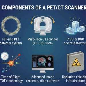
PET CT Scan Machine Components
Common PET CT Scanner Models Used in India
Widely installed systems include:
-
Siemens Biograph Horizon / Vision
-
GE Discovery IQ / MI DR
-
Philips Vereos PET/CT
-
United Imaging uMI Series
Most Indian diagnostic centers operate 16-slice to 64-slice PET CT configurations.
Patient scan cost (approx.)
-
₹18,000 – ₹30,000 per scan
-
Varies by tracer, city, and clinical protocol
PET CT machine price (India)
-
New system: ₹12 crore – ₹25 crore
-
Refurbished system: ₹4 crore – ₹9 crore
Pricing depends on:
-
Slice count
-
Detector technology
-
TOF capability
-
Reconstruction software
-
Service contract duration
PET CT Installation Requirements
Space and Infrastructure
-
Total area: 2,500 – 4,000 sq. ft.
-
Dedicated hot lab
-
Injection room
-
Uptake rooms (shielded)
-
Scan room with lead lining
-
Reporting and control room
Power and HVAC
-
Three-phase stabilized power
-
Dedicated UPS
-
Precision air conditioning (22–24°C)
-
Humidity control
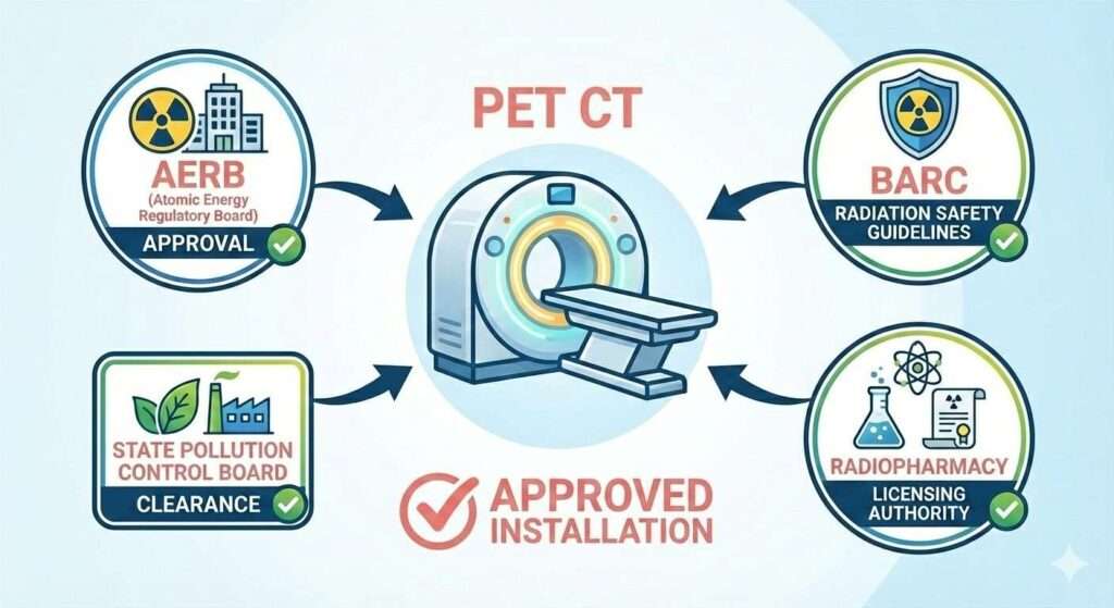
Regulatory and Compliance Requirements in India
PET CT installation requires approvals from:
AERB (Atomic Energy Regulatory Board)
BARC radiation safety guidelines
State Pollution Control Board
Radiopharmacy licensing authority
Staff must include:
Certified Nuclear Medicine Physician
Trained Radiopharmacist
AERB-licensed technologists
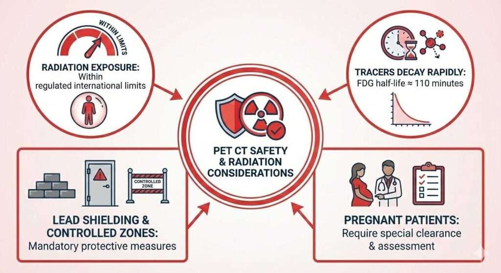
Safety and Radiation Considerations
-
PET CT radiation exposure is within regulated international limits
-
Tracers decay rapidly (FDG half-life ≈ 110 minutes)
-
Lead shielding and controlled zones are mandatory
-
Pregnant patients require special clearance
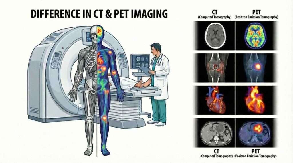
CT shows structure. PET CT shows disease activity.
PET CT vs CT Scan: Key Differences

| Parameter | PET CT | CT Scan |
|---|---|---|
| Detects metabolism | Yes | No |
| Anatomical imaging | Yes | Yes |
| Cancer staging | Excellent | Limited |
| Early disease detection | Very high | Moderate |
| Functional imaging | Yes | No |
Frequently Asked Questions (FAQs)
What is a PET CT scan in simple words?
Why is PET CT mainly used for cancer?
Can PET CT detect early cancer?
How long does a PET CT scan take?
Is PET CT better than CT scan?
Summary
A PET CT scan is a high-precision diagnostic imaging system that combines metabolic and anatomical data in a single examination.
It plays a critical role in oncology, cardiology, and neurology, especially for early detection and treatment monitoring.
For hospitals and diagnostic centers, PET CT represents a strategic long-term imaging investment, requiring regulatory compliance, trained personnel, and advanced infrastructure—but offering unmatched diagnostic accuracy in return.