
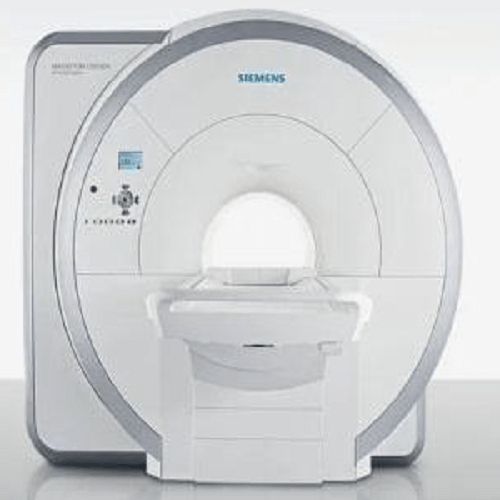
Magnetic Resonance Imaging (MRI) is one of the most powerful diagnostic tools in modern healthcare. When people ask “What is the most common use of MRI?”, the clear and most widely accepted answer is imaging the brain and spinal cord to diagnose neurological conditions.
MRI is most commonly used to examine soft tissues, especially in the brain, spine, and nervous system, because it provides extremely detailed images without using ionizing radiation. This makes MRI safer and more precise than many other imaging techniques for neurological evaluation.
Why Is MRI Most Commonly Used for Brain and Spine Imaging?
MRI scans are especially effective for detecting abnormalities in the central nervous system. The most common clinical uses include:
- Diagnosing brain tumors
- Detecting stroke and brain hemorrhage
- Identifying multiple sclerosis (MS)
- Evaluating spinal disc herniation
- Assessing nerve compression and spinal cord injuries
Because MRI offers high-resolution images of soft tissue, it allows doctors to identify conditions at an early stage, often before symptoms become severe.
How MRI Helps in Neurological Diagnosis
The most common use of MRI is to visualize changes in brain and spinal tissue that cannot be seen clearly on X-rays or CT scans. MRI helps clinicians:
- Measure brain structure and function
- Detect inflammation or infection
- Monitor disease progression
- Plan surgeries with high precision
Advanced MRI techniques, such as functional MRI (fMRI) and diffusion MRI, further enhance diagnosis and treatment planning.
Other Common Uses of MRI (Secondary Applications)
While neurological imaging is the most common use, MRI is also widely used for:
- Joint and musculoskeletal imaging (knee, shoulder, spine)
- Cardiac MRI to assess heart structure
- Abdominal and pelvic imaging
- Oncology for tumor detection and staging
However, brain and spinal MRI remain the top and most frequent applications worldwide.
Why MRI Is Preferred Over Other Imaging Methods
MRI is often chosen because it:
- Does not use radiation
- Provides superior soft-tissue contrast
- Delivers accurate and reproducible results
- Is safe for repeated follow-up scans
These advantages explain why MRI is most commonly used for neurological and spinal diagnostics
- What is the most common use of MRI?
The most common use of MRI is brain and spinal cord imaging. Doctors use MRI scans to detect neurological conditions such as brain tumors, stroke, multiple sclerosis, spinal disc problems, and nerve compression with high accuracy.
2 .Why is MRI preferred for brain scans?
MRI is preferred for brain scans because it provides detailed images of soft tissues without using radiation. It can detect small abnormalities in the brain and spinal cord that may not be visible on CT scans or X-rays.
3. Is MRI safe for repeated use?
Yes, MRI is considered very safe for repeated use because it does not use ionizing radiation. This makes it ideal for long-term monitoring of chronic conditions like brain tumors, spinal disorders, and neurological diseases.
4 .How long does an MRI scan usually take?
An MRI scan typically takes 20 to 45 minutes, depending on the body part being examined and the type of MRI study required. Advanced MRI systems may reduce scan time further.
5. What is the difference between MRI and CT scan?
MRI uses magnetic fields and radio waves, while CT scans use X-ray radiation. MRI is better for soft tissues like the brain, nerves, and muscles, whereas CT scans are commonly used for bones and emergency trauma cases.